Thursday, March 27, 2014

Designer Bags and Designer Chromosomes

      Imagine your pet carrying a genome assembled by a group of scientists. This situation probably sounds like the beginning of a science fiction movie, but it may become a reality. A new report in Science describes the creation of a synthetic chromosome in baker's yeast (Saccharomyces cerevisiae).

      Jef Boeke's group at New York University generated a synthetic version of chromosome 3, one of the smallest chromosomes in the genome of baker's yeast. In creating this chromosome, they eliminated transposons (DNA sequences
that have the ability to move around the genome) and introns (non-coding
regions of DNA). Boeke's group also designed custom DNA sequences
Electron micrograph of human chromosomes.
Courtesy: Andrew Syred, York University, Canada


that they incorporated into the yeast chromosome with the aid of homologous recombination, which involves the exchange of native yeast DNA sequences for the custom-made counterparts. Yeast cells carrying this chromosome were able to mate and behave normally, which indicates the artificial DNA is not deleterious to the cells. Additionally, artificial DNA incorporated into this synthetic chromosome allowed yeast cells to undergo genetic recombination, which can add or delete specific segments of DNA.

      Craig Venter announced his team's creation of the first synthetic bacterium in 2010, and this innovation quickly grasped the attention of the public. With the advent of these synthetic organisms, scientists have claimed that the formulation of vaccines, medications, and biofuels can be less expensive and time-consuming. It may take several years for scientists to engineer these organisms, but this possibility opens the door for additional advantages of these systems to be explored.

Tuesday, March 25, 2014

The Nose Knows More Than We Think

         The intoxating smell of bacon permeates the kitchen. The spring flowers bloom and envelop the atmosphere with their natural scents. Smoke from a nearby bonfire on the beach signals the beginning of summer. These stimuli are sensed by the nose, which houses specialized structures that sense odorants (molecules that produce smells) and relay information to the brain through their association with brain cells. Our noses are able to detect a wide range of smells, and several publications have estimated that the human nose can detect 10,000 different scents.

          However, a report in Science estimates that one trillion scents can be detected by the human nose. Researchers at Rockfeller University developed a set of criteria that allowed for the generation of various odor combinations, which they provided to subjects. The ability of the subjects to discriminate between different odor mixtures was measured, and the quantifications from these experiments were used to calculate their estimate.

          This article noted that one trillion is the lower limit of odors our noses can discern, meaning that the actual number may be much higher. This piece of news intrigued me and led me to think of physical properties that confer this capability. How many different olfactory receptors (proteins that bind odor molecules and facilitate the transfer of odor information to the brain) do we have? Do olfactory neurons, the cells through which olfactory receptors transmit signals, function as small or large groups? How would this activity be controlled? Our olfactory systems may be more complex than we previously believed.

          Furthermore, it will be interesting to determine the mechanism through which olfactory receptors process different odor molecules to allow for the brain to distinguish smells. Studying the means by which this sensory information is sorted and interpreted by the brain will also provide more insight into the development of this amazing ability. Until then, happy smelling!

The scent of a rose is composed of 275 odor components (Bushdid et al., Science 2014).
Courtesy: http://www.varietybackyard.net/ho-to-deal-with-some-common-diseases-of-roses/


Sunday, March 23, 2014

Does Lupus Provide the Basis for the Next HIV Vaccine?

        The immune system allows our bodies to maintain their integrity by eliminating deleterious foreign agents. The immune cells that mediate this process are highly effective and specific, contributing to the powerful nature of the immune response. However, the deregulation of immune cell activity has severe consequences. For instance, lupus is a disorder that stems from the production of self-reactive antibodies (antibodies that recognize the body's proteins). In normal individuals, immune cells that produce self-reactive antibodies are eliminated. However, individuals affected with lupus fail to clear these cells, resulting in autoimmune responses throughout the body that result in widespread inflammation, pain, and tissue destruction.

         Though the damage inflicted by lupus is extensive, one facet of the disease has been highlighted as a possible treatment option for human immunodeficiency virus (HIV) infections, which kill immune cells and dampen the immune response. Through studies of a young woman affected by both lupus and HIV, researchers at Duke University identified broadly HIV-1-neutralizing antibodies (BnAbs). These antibodies were considered unique due to their ability to recognize HIV proteins, and their presence in the woman may have alleviated the detrimental effects of HIV infection. Wonder and amazement filled many people upon the reception of this news, and some have proposed that further studies of these antibodies may lead to the formulation of new HIV vaccines.

         The findings of this study shed new light on lupus and the process that immune cells utilize to produce antibodies, but many questions remain unanswered. First, it is unclear whether BnAb production is dependent on the production of self-reactive antibodies. The authors of the study observed that BnAbs can bind to human proteins and double-stranded DNA, which suggested that BnAbs were self-reactive. This result highlighted a similarity between BnAbs and antibodies found in lupus patients, leading Duke researchers to postulate that BnAbs and self-reactive antibodies are produced by the same population of immune cells. Second, a normal immune system eliminates cells that produce BnAbs, and it is unknown whether the immune system can be coaxed into producing these antibodies safely. Stimulating a person's immune system to generate these antibodies may also encourage the production of self-reactive antibodies, which will be a dangerous side effect. Thus, more work is required to understand how these antibodies are created and the conditions that favor their production. Scientists must also consider the high mutation rate of the HIV virus, so major hurdles obstruct the possibility that this study will lead to a functional HIV vaccine.


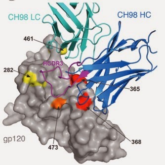
Diagram depicting CH98 (green and blue), a broadly-reactive antibody, bound to the HIV protein gp120 (gray).

Courtesy: Bonsignori et al., The Journal of Clinical Investigation 2014

Friday, March 21, 2014

The Funding Conundrum

          Reused pipette tips were once a odd occurrence in the lab, but they're increasingly more common. The eppendorf tube quality, which was once adequate, are now replaced with shoddy counterparts that fail to close properly and are prone to nail chipping. The purchase of an essential antibody for that dream experiment is now arduously negotiated with the principal investigator, though this practice was virtually nonexistent several years ago. These are all measures that have been taken by labs to cut costs, and unfortunately more drastic steps may quickly follow due to significant funding cuts at the National Institutes of Health (NIH).

          The NIH is the major funding agency for biomedical research, and principal investigators rely greatly on NIH grants to support their research projects and staff. The sequester of 2013 resulted in a
5.5% NIH budget decrease to $29.15 million, which greatly reduced the number of funded research grant proposals in 2013 by roughly 650. The approval rate for grant applications in 2013 was estimated to be 14%, exponentially decreasing the chances that a grant application will be funded. Thus, a proportion of researchers looking to renew their grants were denied and many early career scientists requesting financial support for their projects were turned away.
 
         The scientific community is wading through dark times as a result of this drop in funding. Labs are closing left and right, while those that remain are forced to cut down on resources that are essential for their research. For example, a lab heavily dependent on microscopy for its projects may be forced to decrease microscopy hours. This would negatively impact research quality and progress. Postdoctoral researchers planning to apply for faculty positions are now forced to rethink their options as the possibility of reaching that goal dwindles, and graduate students with the same aspirations are facing the same grim reality.

         Scientific research faces an uncertain future. No substantial remedy for the NIH funding situation has been proposed, and it is possible that more cuts will be made to the budget. The ramifications of this dilemma ripple through the research hierarchy, and talented scientists that may have successfully led their own labs are increasingly drawn toward other career paths. Though the government struggles to maintain our country's infrastructure, officials need to ensure that biomedical research is sufficiently funded. The etiologies of many developmental disorders and diseases remain unclear, and treatment alternatives as well as prophylactic therapy for these maladies depend on the work of research scientists. It is our hope that Congress keeps these considerations in mind as we move forward.  

Friday, March 14, 2014

Happy Pi Day!

             Combine the world's favorite irrational number and a sweet confection of the same name and what do you get? Pi Day! Today is March 14, or 3/14, so I'll shed some light on the history of this popular constant.

            The origins of pi (or 3.14159...) lie in the detailed calculations of the Babylonians and Egyptians, who noted that an approximate ratio of 3:1 described the relationship between a circle's circumference and diameter. The first approximation of pi was achieved by the Greek scholar Archimedes. Unlike his unsuccessful predecessors, he chose to utilize the perimeters of polygons to estimate a value for pi. This involved inscription and circumscription of hexagons followed by a doubling of the sides four times to generate a 96-sided polygon. This significant breakthrough sparked the curiosity of a French mathematician named Françiose Viéte. He adapted the methodology of Archimedes to calculate a pi value of 3.1415926535, the first numerical approximation. Several other gifted mathematicians followed suit and estimated the value of pi to a total of 35 digits. Ludolph Van Ceulen of Germany devoted his life to elucidating the full value of pi and successfully derived the 35-digit value. The symbol for pi (π) which we hold so dear, was introduced by Leonhard Euler in 1737.

          Circles can represent a multitude of ideas. For example, they are believed to symbolize eternity, or the absence of a beginning or end. As the significance of the circle will continue to live on in cultures across the world, the concept of pi will certainly follow in the same footsteps.

courtesy: www.rebloggy.com

Wednesday, March 12, 2014

Embryonic Development of C. elegans

      In about a month, I will be educating kids about the link between genes and behavior at Science Saturday! I belong to a group of students and postdoctoral fellows that will construct booths to show kids that science is fun. And in the process, we will expose them to a multitude of scientific areas. This is an event put together by two science outreach groups at Rockefeller University and it's going to be amazing.

      For my booth, I will present normal and abnormal strains of a small, transparent worm called Caenorhabditis elegans, or C. elegans. This worm is 1 millimeter in length and can be found in common outdoor areas such as compost heaps and garden soil. Due to its short generation time and relatively small genome, this worm has become popular for genetic studies mainly through the work of Nobel laureate Sydney Brenner.

      By training I'm a developmental biologist, so visualizing the metamorphosis of a fertilized egg into a functional organism is quite breathtaking. Enjoy the video below of C. elegans development! How a fertilized egg develops into a hatching worm within twelve hours is nothing short of awesome!

  )
  courtesy: Hymanlab

Thursday, March 6, 2014

Is Tongue-Rolling a Genetic Trait?

          "How many people in this room can roll their tongue?" This question was posed to my ninth grade biology class as an introduction to the subject of genetic traits. I was taught that tongue-rolling, the ability to elevate the lateral edges of the tongue, was a genetic trait; thus, if you were able to roll your tongue, at least one of your parents would be able to do the same. This example was widely utilized in schools to introduce children to the concept of genetic inheritance, but can this trait be  explained by a single gene?

          Several studies in the past 70 years have set out to answer this question. Alfred Sturtevant, of later Drosophila melanogaster (fruit fly) genetics fame, was scintillated by this question and conducted a small survey on those able to roll their tongue (rollers) and those that could not roll their tongue (non-rollers) within families. Before embarking on this project, he hypothesized that tongue-rolling was determined by a single gene with two copies (one inherited from each parent). During his studies, he found that tongue-rolling parents often had children with the same trait. However, he also discovered that parents without the tongue-rolling trait occasionally had children that rolled their tongues, albeit at a lower frequency than parents with the trait. Philip Matlock conducted a similar study in identical twins across a large age range and noted many cases in which only one twin was a tongue roller. The data collected by Sturtevant, Matlock, and others suggest that tongue-rolling cannot be attributed to one gene. In fact, this trait can also be learned because in 1951, Taku Komai's analysis of Japanese children described the increase in tongue-roller frequency from 54% in children from ages 6-7 to 76% in children at age 12.

Courtesy: fakescience.org


        Since there is overwhelming evidence that tongue-rolling is not controlled by a single gene, what are the other possibilities? One is that tongue-rolling is determined by multiple genes, so it would be quite complex to predict who possesses this trait since the identity and quantity of genes responsible for this trait are unknown. Another is that this trait requires an environmental component, and this would be compatible with the observation that non-rollers can learn to roll their tongues. In light of this data, it is inaccurate to state that tongue-rolling is a simple genetic trait. Therefore, it would be more wise for classrooms to revert to pea color and shape as examples of Mendelian inheritance.

Wednesday, March 5, 2014

The Battle of the Sexes: Revisited

     This divide between the sexes is attributed to sex hormones, namely estrogen and testosterone. Both hormones are present in men and women, but testosterone predominates in males while estrogen predominates in females. Several organs in the body contain cells whose behavior is modulated by biological sex. For example, female muscle stem cells were found to regenerate muscle cells more efficiently than their male counterparts in a mouse model of muscular dystrophy. Though sex can influence cellular behavior in multiple organs, cells within organs that are not known to present sex-specific differences are thought to function similarly in both men and women.

     However, a recent report in Nature challenges this notion. Studies in the Morrison lab at the University of Michigan have focused on identifying sex-specific differences in cell populations that are seemingly unresponsive to sex hormones, and hematopoietic stem cells, or HSCs, first grabbed the lab’s attention. Hematopoietic stem cells reside in the bone marrow and divide to generate red blood cells, which carry oxygen to tissues throughout the body, and immune cells, which recognize and destroy bacteria, viruses, and parasites. These cells often lie dormant until stimulated to divide by external cues, and there has been no previous evidence that these cells exhibit sex-related differences. Surprisingly, data from the Morrison group suggest that HSC behavior is not uniform between males and females. Though HSC number and frequency in male and female mice were comparable, increased HSC division was observed in female mice. Interestingly, HSC division was induced by administration of estrogen to both male and female mice, and this phenomenon did not occur in other bone marrow cell populations or in response to testosterone. HSC number, along with red blood cell and immune cell numbers, were increased in pregnant females compared to non-pregnant females. This effect was abolished when estrogen receptor function was compromised, leading to the theory that female HSCs respond to estrogen and that estrogen can regulate HSC division to accommodate the increased need for oxygen in pregnant females. 


    If these data mirror HSC activity in humans, there are several implications. First, the ability of HSCs to respond to estrogen may be relevant for the health of pregnant mothers and their unborn children. Estrogen levels among women can vary, so those with lower estrogen levels may be predisposed to more physical stresses during pregnancy. Second, there is now the possibility that sex hormones can modulate the onset of blood disorders and other diseases. For example, cytopenias, diseases in which immune cell numbers are greatly reduced, tend to be more common in men. Thus, reduced HSC proliferation in men might contribute to the persistence of these disorders. Third, sex may be an additional variable to consider in the field of personalized medicine, which involves the administration of therapy tailored to an individual’s genetic makeup. In this case, it will be imperative to note the individual’s sex in order to uncover contributing factors to disease and to predict patient prognosis. 


    Evolution allows organisms to meet metabolic needs during fluctuations in nutrient availability or changes in physical state (such as pregnancy). It is remarkable that nature may have designed a system that enables HSCs to divide in response to a sex hormone heavily involved in the reproductive process, and new insights provided by future studies on this topic will be of great interest. It will also be fascinating to discover other stem cell populations that function in a similar fashion. Until then, it is comforting to think that womanhood just got a lot better!